Signs of retinal
detachment

Learn to identify the warning signs because early treatment will greatly improve your chances of a positive outcome.
The retina is a thin layer of nerve tissue that lines the wall at the back of the eye, often described as being like the film in a camera. As rays of light pass through our pupils onto the retina, the retina transmits electrical impulses via the optic nerve to our brain. This allows our brain to interpret and make sense of the images around us.
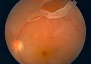
Retinal tear and detachment is a serious condition that occurs as a result of shrinkage of the vitreous – a gel-like material that fills the inside of the eye. This can tug on the retina and cause it to tear. Fluid from the vitreous can then enter through the tear and separate the retina from the back of the eye, causing retinal detachment. If left untreated, retinal detachment can result in permanent loss of vision.

Shrinkage of the vitreous is a condition that usually occurs with age, but it can also be precipitated by trauma, myopia, eye surgeries and inflammations in the eye. There are a number of warning signs which can indicate retinal detachment. They include:
- Sudden onset of dark spots (floaters)
- Sudden flashes of light
- Blurred vision or dark shadows in your field of vision

There are different retinal detachment treatment in Singapore and they include:
Thermal barrier laser photocoagulation
This can be used in those who are diagnosed very early with very limited detachment.
Pneumatic retinopexy
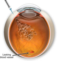
Suitable for those who have retinal breaks at the top of the retina. A gas bubble is injected into the back part of the eye to flatten out the detached retina. Laser photocoagulation is then performed to seal the breaks in the retina.
Scleral buckle
A silicone band is sutured around the sclera (white of the eye) to indent the sclera over the area where the retinal break is. Cryopexy (freezing) is then used to seal the breaks. This band is not visible from the outside of the eye.
Vitrectomy
Involves cutting away the vitreous to relieve traction on the retina, laser photocoagulation to surround the retinal breaks and injection of gas into the eye whilst waiting for the laser to scar and seal the breaks. The individual needs to assume a face down posture for 2 weeks post-surgery.
For people with otherwise normal eyes, the lifetime risk of retinal detachment is one in every 300. This risk increases to one in every 20 for people with high myopia. If you have experienced retinal detachment in one eye, you have a 20% risk of experiencing the same condition in your other eye.
Please visit Eye Max Centre to learn more about retinal tear treatment or contact us at manager@eyemax.sg or +65 6694 1000 to schedule an appointment today.

