VITRECTOMY &
SCLERAL BUCKLE
Retinal detachment is a serious condition that requires immediate treatment. If it is not treated right away, more of the retina may detach, increasing the risk of irreversible vision loss or blindness.
Symptoms of retinal detachment include a sudden appearance of floaters, flashes or a dark shadow moving across your vision. The condition can be caused by injury or by structural changes in the eye over time.
Vitrectomy and scleral buckle are 2 of the key treatments for retinal detachment – most retinas can be successfully reattached if treated early.
VITRECTOMY
A vitrectomy is a surgical treatment used to treat a variety of disorders such as retinal detachments, advanced diabetic eye disease, epiretinal membranes, macular holes, and so on. It is performed in an operating room under local or general anaesthesia.
Three extremely small incisions are made in the white of the eye (sclera) during a vitrectomy. Microsurgical instruments are then placed via these incisions into the eye to remove the vitreous gel. The vitreous gel is not necessary for the proper functioning of the eye and does not need to be refilled.
Following the removal of the vitreous gel, various operations such as laser therapy, freezing (cryotherapy), membrane peeling, and so on may be required as part of the treatment.
At the conclusion of surgery, your eye surgeon will determine whether a synthetic gas or synthetic oil (silicone oil) should be used to fill the eye. The choice will be influenced by the ailment being treated. Synthetic gases fade away over a period of 2 weeks to 2 months, and are replaced by fluid produced naturally by the eye. Silicone oil, on the other hand, will necessitate a second surgery to remove.
The recovery time from the procedure itself can be several weeks. However, with the small incision sutureless vitrectomy approach, post-operative recovery is easier than before because the smaller wounds cause less discomfort and heal faster.

SCLERAL BUCKLE
Scleral buckle is a surgical procedure used to treat a detached retina. It is performed in an operating room under local or general anaesthesia.
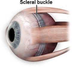
The scleral buckle is a silicone band or sponge sewed onto the eye’s outer wall (sclera), beneath the eye muscles. The buckle is fastened in such a way that it supports the retinal tear that is causing the retinal detachment. The buckle pushes the eyewall inwards, generating an indent in the eyeball that relieves the abnormal internal pressure of the vitreous gel on the retina. It also allows the detached retina to come together with the eyewall and reattach itself. To seal the tear’s edges, laser treatment or freezing (cryotherapy) of the retina is used.
The recovery time from the procedure can take several weeks.

