
Is Retinal Detachment Surgery Considered a Major Surgery?
Summary:
- Retinal detachment surgery is major surgery — a detached retina causes irreversible vision loss if untreated, and repair requires one of three procedures: vitrectomy (TPPV), scleral buckle, or gas/silicone oil placement.
- TPPV, the most common method, removes vitreous gel for a clear surgical view of the detachment.
- Recovery takes 1 to 3 months, often requiring a face-down posture.
- Sudden floaters, flashes, or a dark curtain over vision need urgent same-day review.
- Retina reattachment (anatomical success) doesn’t guarantee full vision recovery, especially if the macula was involved.
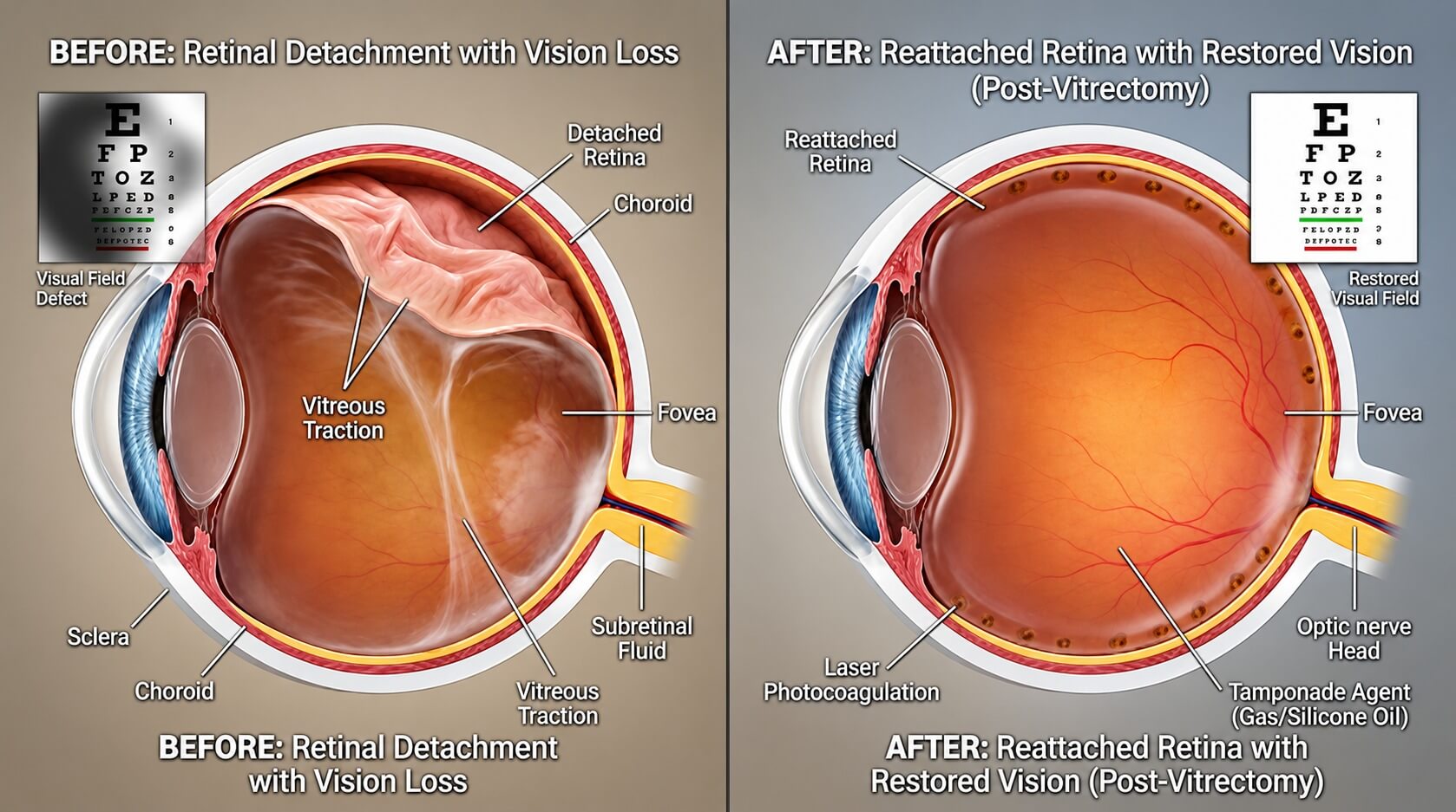
Retinal Tear vs. Retinal Detachment: What’s the Difference?
Retinal detachment surgery is considered a major surgery due to the urgency of reattaching the retina, as this light-sensitive layer is responsible for our vision. If timely intervention is not taken, this can affect the post-surgical visual outcome, as any vision loss from a detached retina is irreversible.
A retinal tear or hole differs from a retinal detachment as it can be repaired with a simple laser, whereas a detachment requires surgical intervention to reattach the retina. A retinopexy laser is applied around the retinal tear or hole to create small adhesive scars that seal the retina to the underlying tissue, helping to prevent fluid from passing through the break and reducing the risk of retinal detachment. If there is fluid accumulation under the retina, the retina lifts away from the other layers, and this lifting is known as a retinal detachment.
Also Read: Retinal Laser Treatment: Benefits and Risks
Retinal Detachment Surgery Methods
There are several ways to repair a retinal detachment:
- Trans pars plana vitrectomy (TPPV): The most common method to repair a detachment, where vitreous gel in the eye is removed to allow the surgeon to have a better view of the detachment.
- Scleral buckle (SB): A soft band is placed around the eye to “squeeze” it and allow the detachment to seal back to the other layers of the retina.
- Gas or silicone oil (SO) placement: Once the surgeon has successfully repaired the detachment, either gas or silicone oil will be injected into the eye. This internal support agent will help to stabilise the retina after surgery.
Also Read: Retinal Disease Treatment
What Type of Anaesthesia Is Used?
The type of anaesthesia used for the surgery varies for each individual and also depends on the complexity of the detachment. For most people, monitored anaesthesia is used for the surgery. In cases where it is a child, someone with anxiety or a complex detachment, general anaesthesia is the preferred choice. However, recovery with general anaesthesia will be slightly longer as the body has to slowly get rid of the effects of the sedation.
Recovery After Retinal Detachment Surgery: What to Expect
As there may be a gas bubble or silicone oil in the eye, vision will be blurry until such time as the gas bubble is slowly reabsorbed back into the body or the silicone oil is surgically removed. The recovery process ranges from 1 to 3 months. A face-down posture needs to be adopted to allow the support agents to act as an internal “band-aid” on the area of detachment. It is crucial during this phase to avoid engaging in any strenuous activities and to take everything light and easy. Your ophthalmologist will also review you closely to ensure no other post-operative complications occur during this period. If the macula was also detached, vision may not restore to how it was before, as the macula is responsible for sharp, central vision.
What Happens If Surgery Is Delayed?
If there is a delay in surgical intervention, the detachment may get wider over time, and eventually the entire retina will detach, leaving that particular eye completely blind. There is also a possibility that the surgery will get more complex with an increased area of detachment. Visual recovery also declines if the retina has been detached for a prolonged period.
Is Retinal Detachment Surgery Always Successful?
The majority of the time, retinal detachment surgery is able to successfully reattach the retina. However, anatomical success does not equivalate to visual recovery. Anatomical success refers to the retina staying reattached after the surgery, while visual recovery equates to how much vision is functional even with a successful surgery. Even with a successful surgery, some people require further treatment. If silicone oil was injected during the first surgery, it needs to be surgically removed once the retina has stabilised. In some cases, scar tissue may develop during the healing process. As this scar tissue contracts, it can exert traction on the retina, potentially leading to new retinal tears or recurrent retinal detachment. Another surgery is needed to remove this scar tissue to ensure that it does not cause further damage to the healing retina.
Warning Signs You Should Not Ignore
If you experience symptoms suggestive of a tear, it requires urgent review with an ophthalmologist. Firstly, sudden floaters are tiny strands or cobweb-looking things that float in and out of your vision. They may appear black or transparent. These are usually harmless; however, if there is a sudden “shower” of floaters, it could possibly mean that there is a tear due to the vitreous gel pulling away from the retina. Another symptom that could occur with or without floaters is flashes. This happens when the vitreous pulls away from the light-sensitive layer and causes flashes, which can appear like flickering of light or a camera flash and are more noticeable in darker areas or at night. If you notice a dark veil that blocks your vision, it is possible that the retina has already detached further into your vision than you are able to notice it. This is warranted as an emergency, and you should see an eye specialist as soon as you can. If you are unable to, you may also go to an emergency department or urgent care centre. When there is a sudden loss of vision, there is a high chance that the macula, the area responsible for our central vision, has detached from the other layers too. When the macula detaches, it makes the surgery more complex, and the vision may not be as good as before the detachment.
For more information about retinal detachment symptoms and treatment, refer to the National Eye Institute.
Final thoughts
If you are experiencing symptoms like floaters, flashes, a dark curtain or sudden vision changes, seek urgent assessment from an eye specialist. Schedule an appointment with Eye Max Centre at manager@eyemax.sg or +65 6694 1000.
